
A patient with a well-differentiated neuroendocrine tumor has a new liver lesion, and the oncology team wants proof that the lesion expresses somatostatin receptors before selecting a receptor-targeted therapy. The team also requires uptake to be measured on PET for eligibility review. Which tracer and preparation combination should be arranged?
Select an answer to reveal the explanation.
Short Explanation
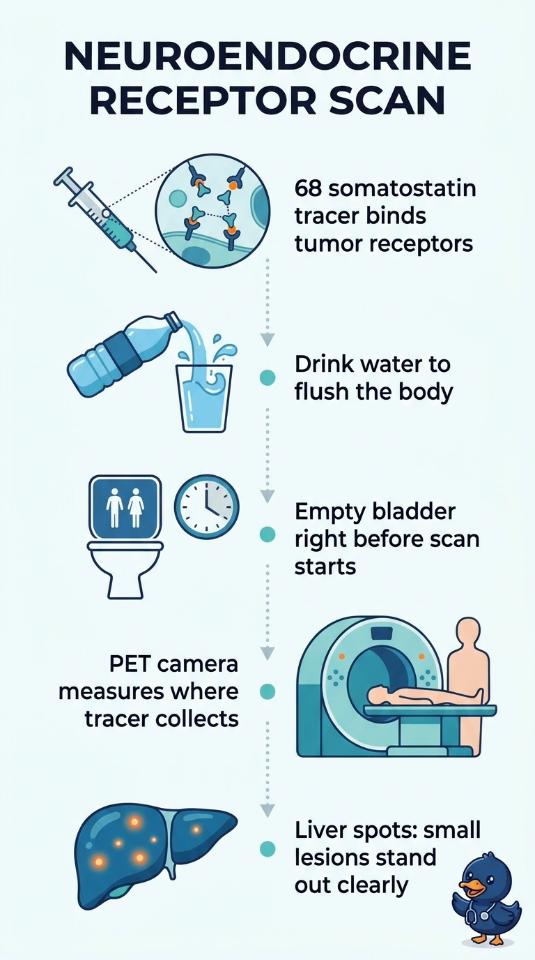
Your clinical question is about receptors, so the tracer has to bind those receptors to answer it. A gallium-68 somatostatin receptor tracer lights up neuroendocrine tissue and still yields PET numbers for the eligibility review. Solid hydration and an empty bladder keep background low so liver lesions read clearly.
Full Explanation
The request combines two requirements that pull in different directions: proof of receptor expression and quantitative PET data for eligibility. A gallium-68 labeled somatostatin receptor tracer binds the receptor these tumors overexpress, so uptake confirms the target and the PET acquisition yields standardized uptake values that can be compared with therapy criteria. Fluorine-18 FDG measures glucose metabolism instead, which is useful in aggressive lymphoma or poorly differentiated disease but does not establish receptor status, and its fasting and glucose check address a different biological pathway. A gallium-68 prostate-specific membrane antigen tracer targets another membrane protein entirely, and its hydration and voiding preparation is sound for background reduction yet irrelevant to receptor expression in a neuroendocrine lesion. Indium-111 pentetreotide does bind somatostatin receptors, yet planar and single-photon emission imaging lack the sensitivity, resolution and quantitation needed for a PET-based eligibility review of small liver lesions. Exam caveat: long-acting somatostatin analog therapy may need coordination with imaging, so check department policy instead of assuming. Practical check: review hydration, renal status and recent receptor therapy before injection.