
An outpatient has a small, well-defined lesion in the right lobe of the liver, and the referring physician wants imaging that shows whether the lesion slowly fills with blood to confirm a cavernous hemangioma. The department stocks sulfur colloid, a hepatobiliary agent, and a red blood cell labeling kit. Which study best demonstrates the blood-pool pattern?
Select an answer to reveal the explanation.
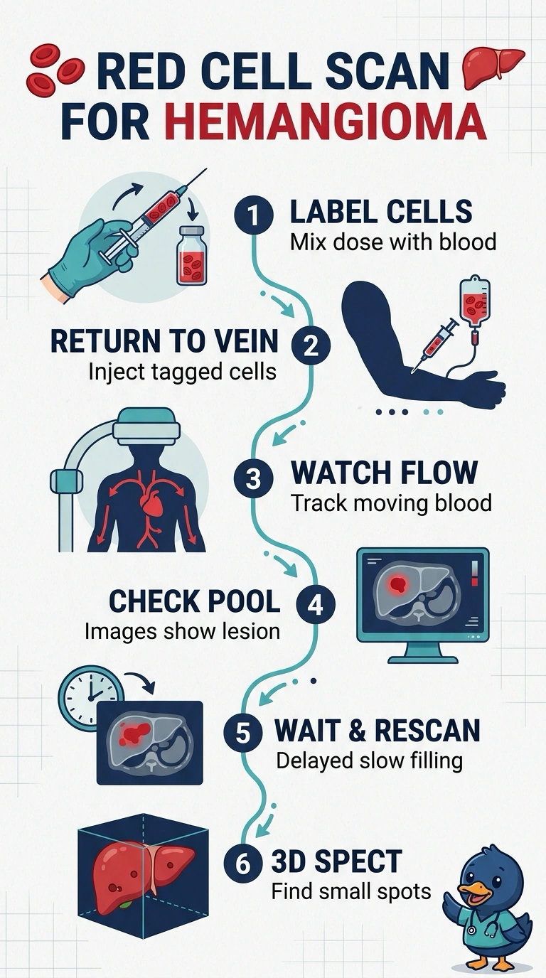
Short Explanation
Slow filling is the signature of a hemangioma, so you need imaging that follows blood pool activity over time, and delayed SPECT is what catches a small lesion. A sulfur colloid scan looks at Kupffer cells, while a hepatobiliary study tracks bile flow instead. It's like glancing at a sponge once versus watching it soak.
Full Explanation
Hemangiomas are slow-flow vascular spaces, so the diagnostic pattern is blood-pool activity that persists or increases on delayed images relative to surrounding liver, with flow images that are normal or mildly prominent; labeling red cells in vitro gives the cleanest pool images, and delayed SPECT adds the depth resolution planar views lack when a small lesion sits near the dome or hides behind liver activity. A sulfur colloid study depends on Kupffer cell phagocytosis, so it can reveal a photopenic defect but never demonstrates filling over time. A hepatobiliary study tracks hepatocyte extraction and biliary excretion, which describes function rather than vascularity. An in vivo red-cell plan restricted to early planar images skips the delayed phase where the gradual fill appears, and in vivo labeling gives lower labeling efficiency than a kit processed outside the body while planar-only imaging can bury a small lesion under overlying activity. The exam caveat is that more than one labeling method appears in practice, so weigh the imaging sequence more heavily than the kit. Practical check: confirm your department's acquisition times for flow, early pool, and delayed images before the patient leaves.