
A patient with severe pulmonary hypertension and a suspected pulmonary embolism is scheduled for a combined ventilation and perfusion study using Tc-99m DTPA aerosol and Tc-99m MAA, and the physician also wants quantitative split-lung values from planar data. Which administration and imaging plan should the technologist follow?
Select an answer to reveal the explanation.
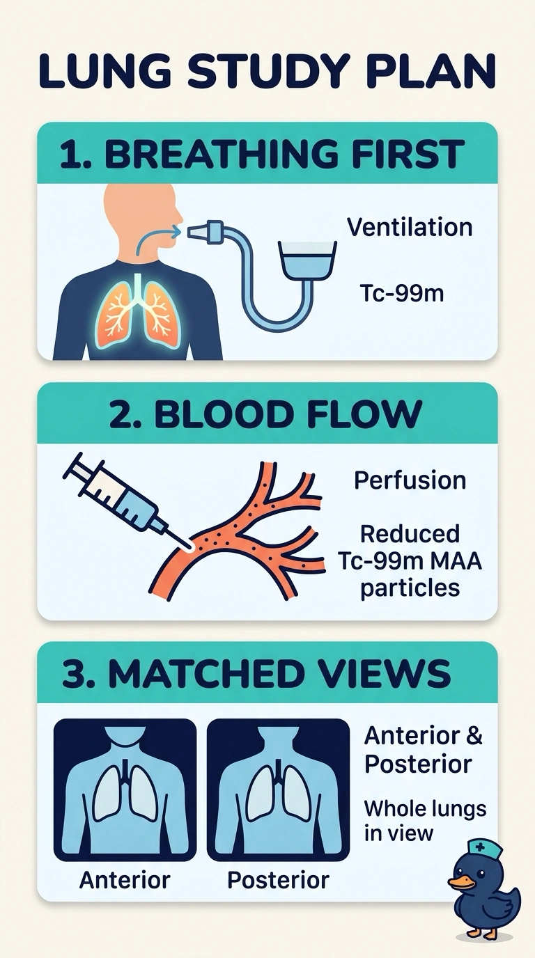
Short Explanation
With high pressure in the lung circulation you inject fewer particles, like sending fewer cars across a weak bridge. Both tracers are Tc-99m, so breathing images must come first or it's impossible to tell the two apart. The split-lung math also needs matched anterior and posterior views.
Full Explanation
Perfusion imaging relies on Tc-99m MAA particles lodging in pulmonary capillaries, and severe pulmonary hypertension leaves little reserve, so the particle count is cut to avoid closing off an excessive share of the bed. Because both the aerosol and the perfusion agent are labeled with Tc-99m, their photopeaks are identical; ventilation is acquired first so trapped MAA never contaminates those images, and a perfusion-first sequence makes the two portions inseparable. Quantitative split-lung values come from matched anterior and posterior whole-lung images, because the geometric mean of those two counts reduces the effect of source depth, so an oblique-only set cannot support the calculation even though oblique views are routine for defect localization. Exam caveat: when a ventilation agent with a different energy is used, the ordering rule can change, so tie your answer to the isotopes named in the stem. Practical check: confirm the physician-approved particle reduction and the required view set before the aerosol is given.