
An 11-year-old has had two episodes of painless, bright-red rectal bleeding, and colonoscopy plus upper endoscopy are normal. The surgeon suspects a Meckel diverticulum with ectopic gastric mucosa. Which Meckel scan plan best demonstrates that ectopic gastric tissue?
Select an answer to reveal the explanation.
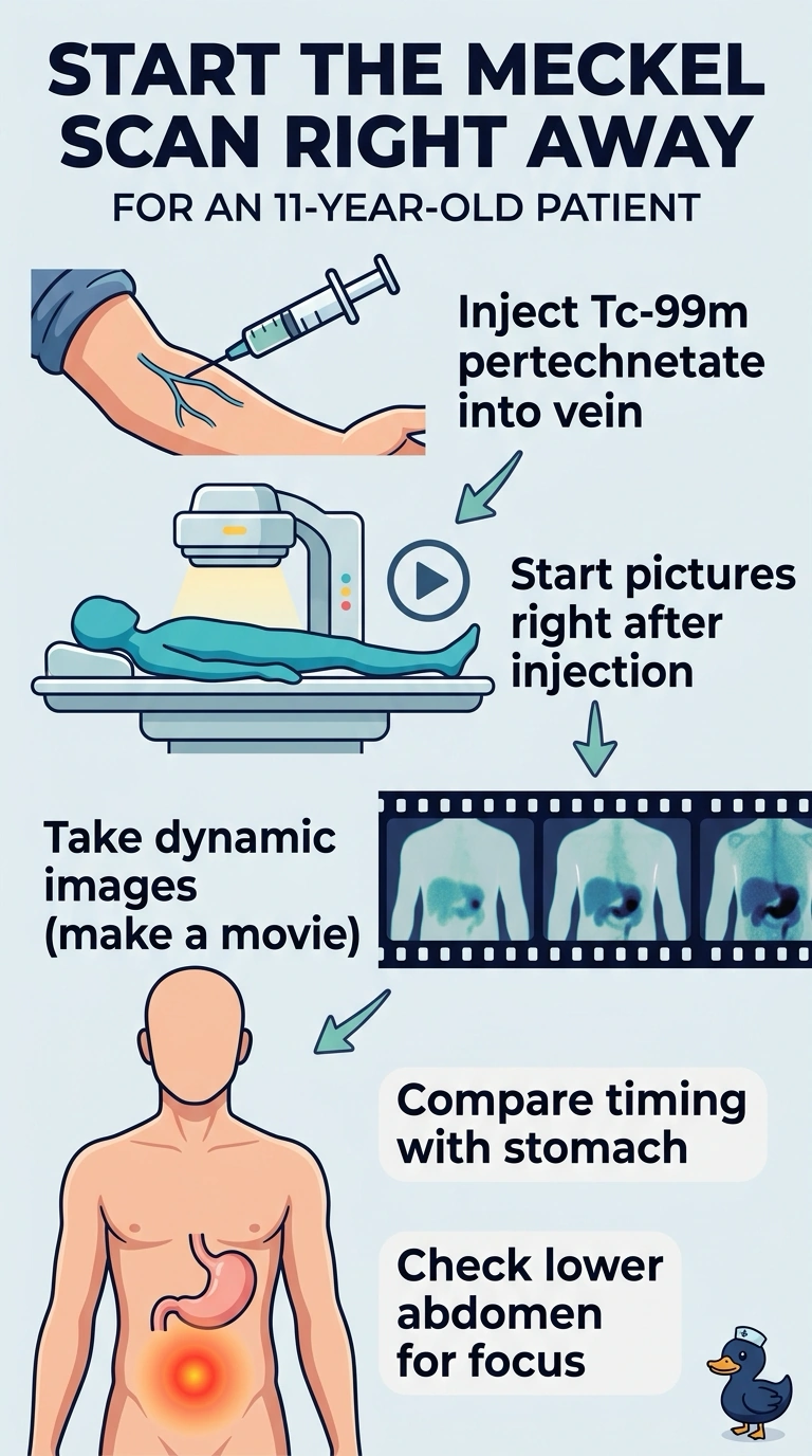
Short Explanation
Pertechnetate is taken up by ectopic gastric tissue, which should light up around the same time as your stomach—like two clocks ticking together. Start dynamic imaging right after injection so you can compare the suspected focus with the stomach; this timing is key to detecting ectopic gastric mucosa.
Full Explanation
Ectopic gastric mucosa in a Meckel diverticulum takes up Tc-99m pertechnetate, as normal gastric mucosa does. Starting dynamic imaging immediately after injection lets the reader follow tracer delivery and recognize a focus that appears in parallel with the stomach; that temporal and spatial comparison supports the diagnosis. Tagged red-cell scintigraphy is a useful method for detecting and localizing active, including intermittent, gastrointestinal bleeding, but it tracks labeled blood rather than gastric mucosa, so it does not demonstrate the tissue suspected here. Tc-99m sulfur colloid can also help localize active bleeding, particularly when bleeding is occurring during imaging, but it likewise does not selectively map ectopic gastric tissue. Immediate static pertechnetate images use the appropriate tracer and include gastric comparison, but a static acquisition does not provide the required dynamic assessment of the focus as uptake develops after injection. A negative Meckel scan does not absolutely exclude a diverticulum: sensitivity depends on functioning ectopic gastric mucosa and can be affected by preparation, medications, or active bleeding. A practical check is to confirm that acquisition begins at injection and that the suspected focus is assessed for timing and intensity relative to the stomach.