
A surgical ward patient has had three episodes of maroon-colored stool over the past day with stable vital signs and a stable hemoglobin. Endoscopy was nondiagnostic, and the surgeon wants the bleeding site localized during the next episode. The department offers labeled red blood cell imaging, a pertechnetate Meckel study, and a sulfur colloid bleed study. Which imaging plan is best for localizing this patient's bleeding?
Select an answer to reveal the explanation.
Short Explanation
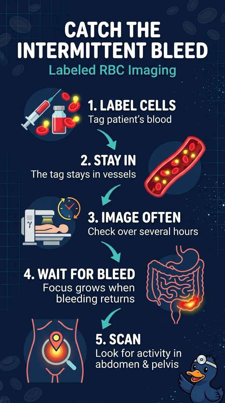
Labeled red blood cell imaging is like keeping a camera ready: the cells stay in your bloodstream for hours, so you can get serial images while waiting for the intermittent bleed. If bleeding resumes, the tracer can build up at the site and help localize it.
Full Explanation
Technetium-labeled red blood cells remain in the vascular space for an extended period, allowing repeat imaging while the technologist waits for bleeding to recur; resumed hemorrhage may appear as a focal collection of activity that increases over time. Because this patient's episodes are hours apart, prolonged imaging is useful. A pertechnetate Meckel study targets ectopic gastric mucosa rather than localizing an active hemorrhage. Limiting labeled red blood cell imaging to the first few minutes would not provide the extended observation needed for an intermittent bleed. Sulfur colloid is rapidly cleared from the blood pool, limiting the period in which it can detect bleeding. Confirm the labeling method in use and arrange repeat imaging as appropriate for the clinical protocol.