
A 62-year-old inpatient has fever after bowel surgery, and CT shows a fluid collection in the right lower quadrant. The surgeon wants an autologous labeled white blood cell study with delayed images before deciding whether to drain it, since the collection sits low in the abdomen where bowel activity can interfere. Which labeling and imaging plan best answers this abscess question?
Select an answer to reveal the explanation.
Short Explanation
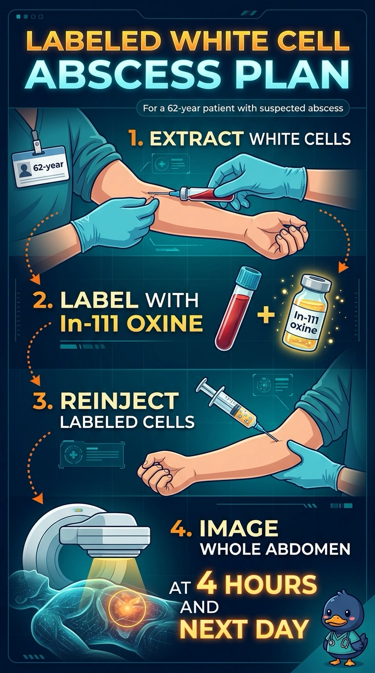
You label your own white cells with In-111 oxine, reinject them, and image your whole abdomen at 4 hours and again the next day. It's like tracking your body's infection-fighters over time, and In-111 does not pass into the bowel the way Tc-99m-labeled cells can, so a low abdominal collection stays visible on the delayed images.
Full Explanation
In-111 oxine labels the patient's own leukocytes, and its 67-hour physical half-life permits imaging the next day. In-111-labeled leukocytes are generally preferred for abdominal or pelvic infection because Tc-99m HMPAO-labeled leukocytes can release activity that is excreted through the hepatobiliary system into the bowel, potentially obscuring a low-abdominal collection. Imaging at about 4 hours and again at about 24 hours provides early and delayed views; uptake and image quality depend on the clinical situation and labeling. Tc-99m HMPAO also labels leukocytes, but its shorter half-life makes next-day imaging unsuitable, and bowel activity may interfere at this site. Sulfur colloid is taken up mainly by reticuloendothelial tissue rather than serving as a label for circulating leukocytes, so it does not answer the question of leukocyte accumulation in the collection. Verify labeling efficiency and follow radiation-safety procedures during reinjection.