
A woman with a palpable upper outer quadrant breast tumor is booked for sentinel node mapping, with injection at 8 AM and transport to surgery at 9:30 AM. The surgeon wants to see the lymphatic channels leaving the primary itself and needs to know how deep the node sits before the incision. Which acquisition plan fits this lymphoscintigraphy study?
Select an answer to reveal the explanation.
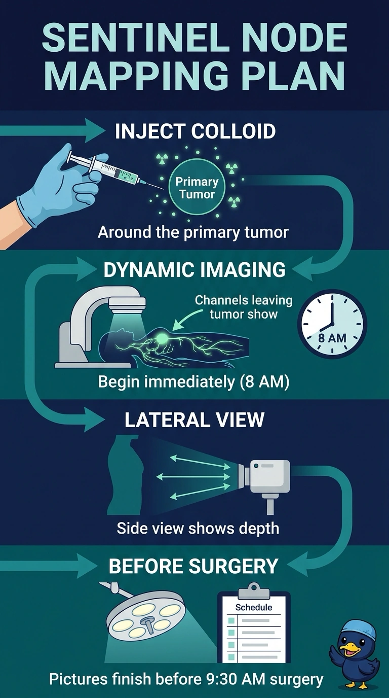
Short Explanation
Your injection site decides which lymphatic channels show up, so a peritumoral injection puts the tracer right where the surgeon is looking. Dynamic imaging immediately after injection captures lymphatic transit before the 9:30 transport, and the lateral view adds depth the front view cannot give you. That's the difference between a street address and a full address that includes the floor number.
Full Explanation
Planning sentinel lymphoscintigraphy means matching three variables to the surgical question: where the tracer goes, when you image, and from which angle. Depositing the colloid around the primary places activity in the lymphatic channels that drain the tumor, which is exactly the drainage this surgeon asked to see; beginning dynamic imaging immediately after injection captures transit while the patient is still in your department, before the transport window, and lateral or oblique views supply the depth dimension that lets the incision be planned in three dimensions instead of two. Delayed-only acquisition fails the timing requirement, because imaging that starts after the patient leaves offers no pre-incision map. Periareolar injection fails the site requirement, since it maps drainage from the areolar plexus, which may share the axillary basin but does not show the channels leaving the primary itself. Anterior-only acquisition fails the depth requirement, giving a hotspot on the chest wall without showing whether it lies superficial or deep. Exam caveat: activity, volume, and exact delay times vary by protocol and by manufacturer guidance, so anchor on site, timing, and projection rather than fixed numbers. Practical check: confirm the marked tumor location, agree the image timing with the surgical team, and obtain at least one lateral view before the patient leaves.