
A clinical AI project outlines tumor boundaries pixel-by-pixel on radiology scans so clinicians can see exact lesion regions. What computer vision task does this describe?
Select an answer to reveal the explanation.
Short Explanation and Infographic
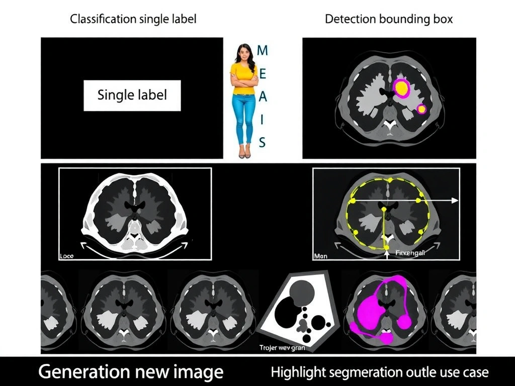
Check this out: if every pixel gets a label—tumor vs healthy tissue—that's segmentation. Think of it like coloring inside the lines of a lesion map, not just sticking a sticky-note box on the page. Object detection gives you rectangles. Classification says "tumor present" for the whole slice. Generation makes brand-new pictures. Your boss wants outline accuracy for surgery planning? That's masks, not boxes. Exam trap: people mix detection and segmentation because both "find" objects—remember density. Detection = where roughly; segmentation = which pixels. Land the takeaway: pixel-wise regions = image segmentation.
Full explanation below image
Full Explanation
Computer vision tasks differ by the granularity of their outputs. Image segmentation predicts a label for every pixel (or a continuous mask), grouping pixels into semantic classes (semantic segmentation) or into individual object instances (instance segmentation). Tumor delineation on medical images is a textbook segmentation use case: clinicians need the spatial extent of abnormal tissue, not merely a flag that something is present or a coarse box around a lesion. Architectures such as U-Net and related encoder–decoder designs are widely used because skip connections preserve fine spatial detail while hierarchical encoders capture broader anatomical context across the scan.
Object detection outputs bounding boxes (or oriented boxes / keypoint sets) with class scores. Boxes are efficient for counting and coarse localization but include background inside the rectangle and omit irregular lesion shapes that matter for volume estimates and treatment margins. Whole-image classification maps an entire scan or patch to one or a few labels; it supports triage workflows ("likely malignant") but provides no contour for measurement or radiation planning. Image generation synthesizes new samples (diffusion models, GANs) and is orthogonal to analyzing an existing diagnostic image for regions of interest—the clinical scenario starts from a real patient scan that must be interpreted, not invented.
In regulated medical settings, segmentation quality is judged with metrics like Dice coefficient, Intersection-over-Union, Hausdorff distance, and clinical review of boundary errors. Preprocessing (windowing, resampling), careful multi-rater annotation protocols, and robustness to domain shift across scanners and protocols all matter in deployment. Teams also monitor false-positive speckles and missed satellite lesions. Memory aid: classification = one label for the picture; detection = box around the thing; segmentation = paint every pixel. When a scenario emphasizes outlining regions pixel-by-pixel on medical images, the correct task name is image segmentation.