
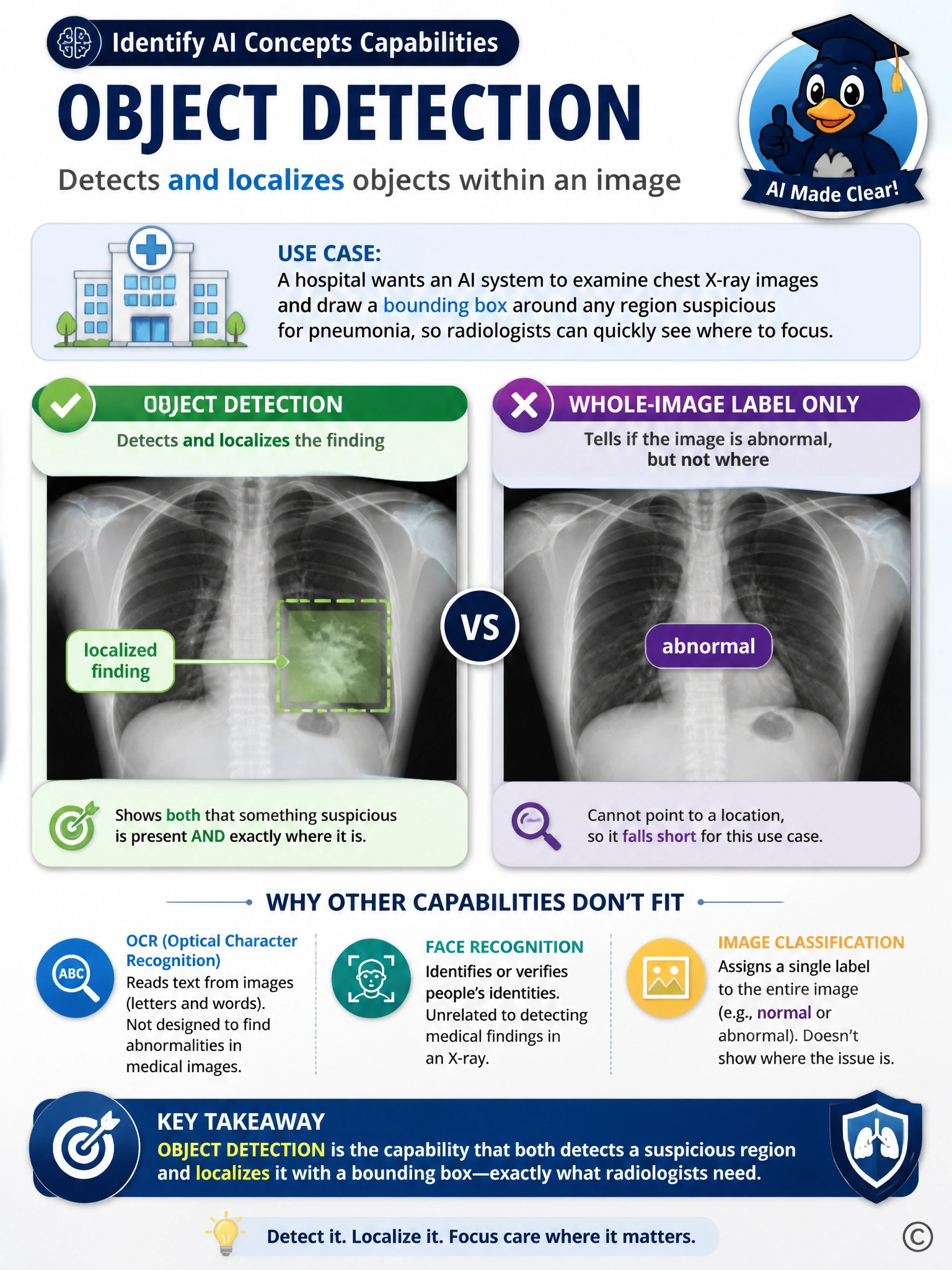
A hospital wants an AI system to examine chest X-ray images and draw a bounding box around any region suspicious for pneumonia, so radiologists can quickly see where to focus. Which computer vision capability directly supports this requirement?
Select an answer to reveal the explanation.
Short Explanation
The hospital wants two things at once: knowing something suspicious is there, and knowing exactly where it sits on the image so a bounding box can be drawn around it. Whole-image labeling can tell you the picture as a whole looks abnormal, but it cannot point to a location, so it falls short of what is being asked. Reading text off an image is a completely different skill aimed at letters and words, not lung tissue. And recognizing faces is built for matching identities, which has nothing to do with spotting a shadow on an X-ray. The capability that both detects and localizes a region within an image, box and all, is the one that matches this radiology use case.
Full Explanation
The correct answer is D. Object detection not only identifies what is present in an image but also locates it by drawing a bounding box around the region of interest, which matches the hospital's need to highlight the suspicious area on the X-ray for the radiologist. Option A is incorrect because image classification assigns a single label to the entire image, such as 'pneumonia present' or 'normal', without indicating where in the image the finding is located, so it cannot draw a bounding box. Option B is incorrect because optical character recognition extracts printed or handwritten text from images, which is irrelevant to identifying a visual abnormality in lung tissue. Option C is incorrect because facial recognition is a specialized technique for identifying or verifying human faces and has no application to interpreting anatomical structures on a radiograph.