
In a sports clinic a 19-year-old soccer player has lateral ankle pain after inversion, the AP and lateral are normal, and the physician suspects syndesmotic widening that requires symmetric joint spaces on a true mortise view for comparison. Which positioning action should the radiographer take to obtain an optimal ankle mortise projection?
Select an answer to reveal the explanation.
Short Explanation
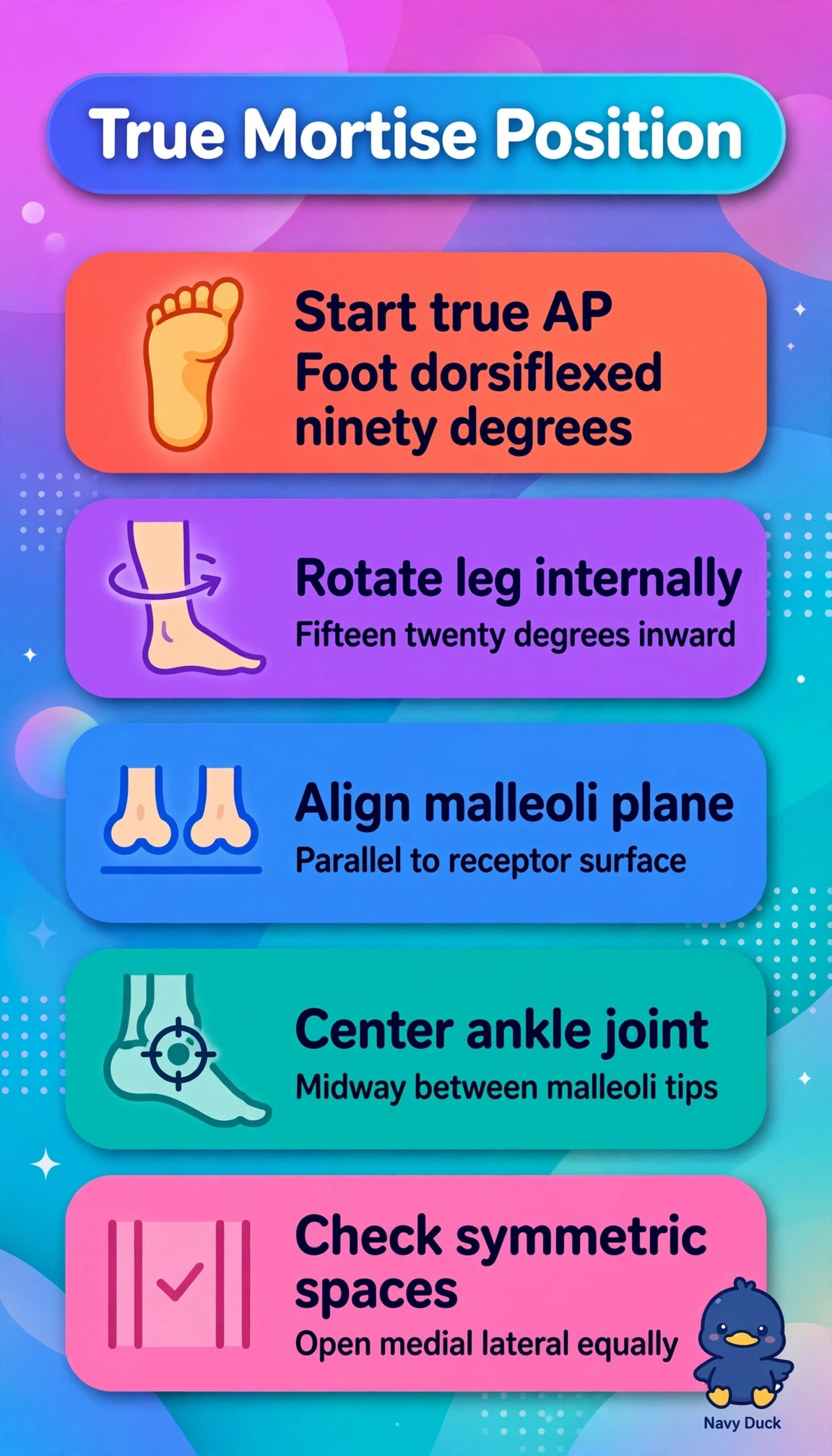
You’re looking for a joint view that can hide unless you angle the ankle just right. Turn the leg and foot slightly inward to open the mortise with symmetric spaces.
Full Explanation
An optimal ankle mortise view requires the intermalleolar plane to be parallel to the image receptor, which makes the medial and lateral joint spaces appear symmetric and open. Internally rotate the entire leg and foot 15 to 20 degrees; this obliquity compensates for normal tibial torsion. Keep the foot dorsiflexed to 90 degrees and center to the joint. Externally rotating the leg and foot reverses the required obliquity, further closes the mortise, and fails to produce symmetry. Dorsiflexing the foot and angling the central ray 15 degrees cephalad without rotating the leg changes the beam angle but does not correct the intermalleolar plane, so the spaces remain asymmetric. Plantarflexing the foot and everting the ankle distorts tibiotalar alignment and imitates a stress maneuver rather than meeting true mortise criteria. Exam caveat: the mortise is an internally rotated AP oblique ankle projection; stress and weight-bearing views have separate indications. Practical check: palpate both malleoli, rotate until they are equidistant from the receptor, dorsiflex the foot to 90 degrees, and confirm that the medial and lateral clear spaces are open.