
A 28-year-old athlete in sports medicine has suspected scaphoid fracture, wrist taped in ulnar deviation, thin part with fine trabeculae to show, and prior images looked flat with magnified oval carpus. Which combination of exposure and geometry best optimizes subject contrast, spatial resolution, and shape distortion for this small-bone study?
Select an answer to reveal the explanation.
Short Explanation
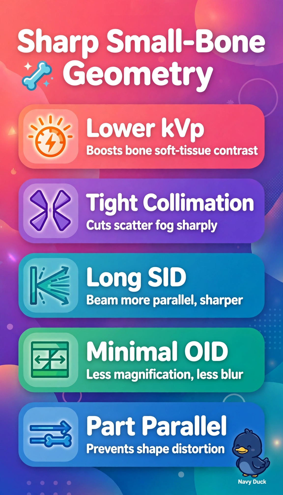
You've got tiny bone detail fighting gray wash and blur, so tightening the beam geometry and contrast wins. Lower kVp boosts subject-contrast while long SID and low OID keep edges sharp, like switching to macro mode for jewelry.
Full Explanation
Subject contrast depends on differential attenuation, so lower kVp widens differences between bone and soft tissue for a thin wrist, while tight collimation limits scatter that would otherwise fog fine trabeculae. Spatial resolution improves with a smaller effective blur from geometry, achieved by long SID that makes the beam more parallel and minimal OID that reduces magnification and penumbra, plus keeping the part parallel prevents shape distortion from unequal magnification. Higher kVp with wide collimation fails by concept because it lowers photoelectric differences and adds scatter despite good geometry, producing flat gray tones. Short SID with increased OID fails by geometry because magnification and focal-spot penumbra increase and degrade spatial resolution despite good kVp. Angling the wrist to the receptor fails by distortion because the projected shape becomes foreshortened and oval despite good contrast and sharpness, misrepresenting carpal anatomy. Exam caveat is that distortion and unsharpness can mimic or hide fracture lines, so always separate contrast factors from geometry factors. Practical check is to palpate that the wrist is flat on the receptor, measure SID at 40 inches, and select the lowest kVp that still penetrates without excessive mAs.