
A 58-year-old with liver metastases is staged for Y-90 microsphere therapy, and angiography shows the treatment catheter will sit in the proper hepatic artery. Nuclear medicine must produce the pretherapy Tc-99m MAA study to estimate the lung shunt fraction before the dose is ordered. Which injection and imaging plan best supports that shunt estimate?
Select an answer to reveal the explanation.
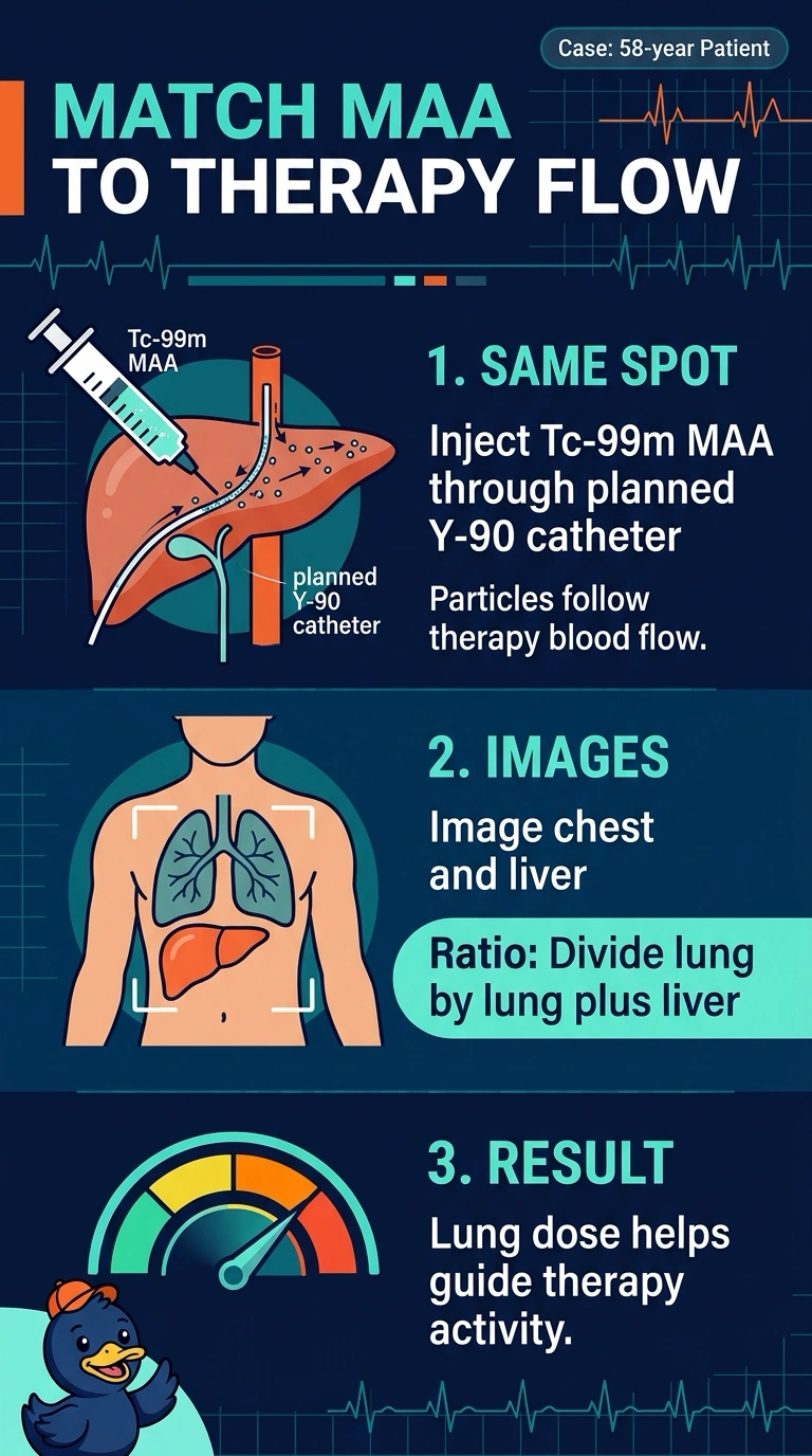
Short Explanation
You want the MAA to take the same road as the Y-90 microspheres, so inject it through your planned treatment catheter position. Then image the chest and liver to estimate the lung shunt fraction—like a test run that shows how much of the dose may travel to the lungs.
Full Explanation
Radioembolization distributes microspheres by arterial flow, so the surrogate should enter the intended vascular territory from the planned treatment catheter position. Tc-99m MAA injected there travels through the hepatic artery and distributes in the liver and tumor beds; particles that pass through hepatic vascular shunts may reach the lungs, which is what the lung shunt fraction evaluates. Imaging both the chest and the liver supplies the numerator and denominator: lung activity is compared with lung plus liver activity, and the resulting estimate helps assess lung dose and guide therapy planning. A peripheral intravenous injection sends MAA to the lungs through the right heart, measuring pulmonary perfusion rather than hepatic arterial shunting, and bypasses the tumor supply. An arterial injection from a different position may distribute particles to different territories, so the estimate may not match the planned delivery. Chest-only imaging does not provide the liver counts needed for the lung shunt fraction. Particle quantities and image timing vary across protocols, so follow the local protocol. Confirm the planned catheter position with the interventional team before drawing up the MAA.