
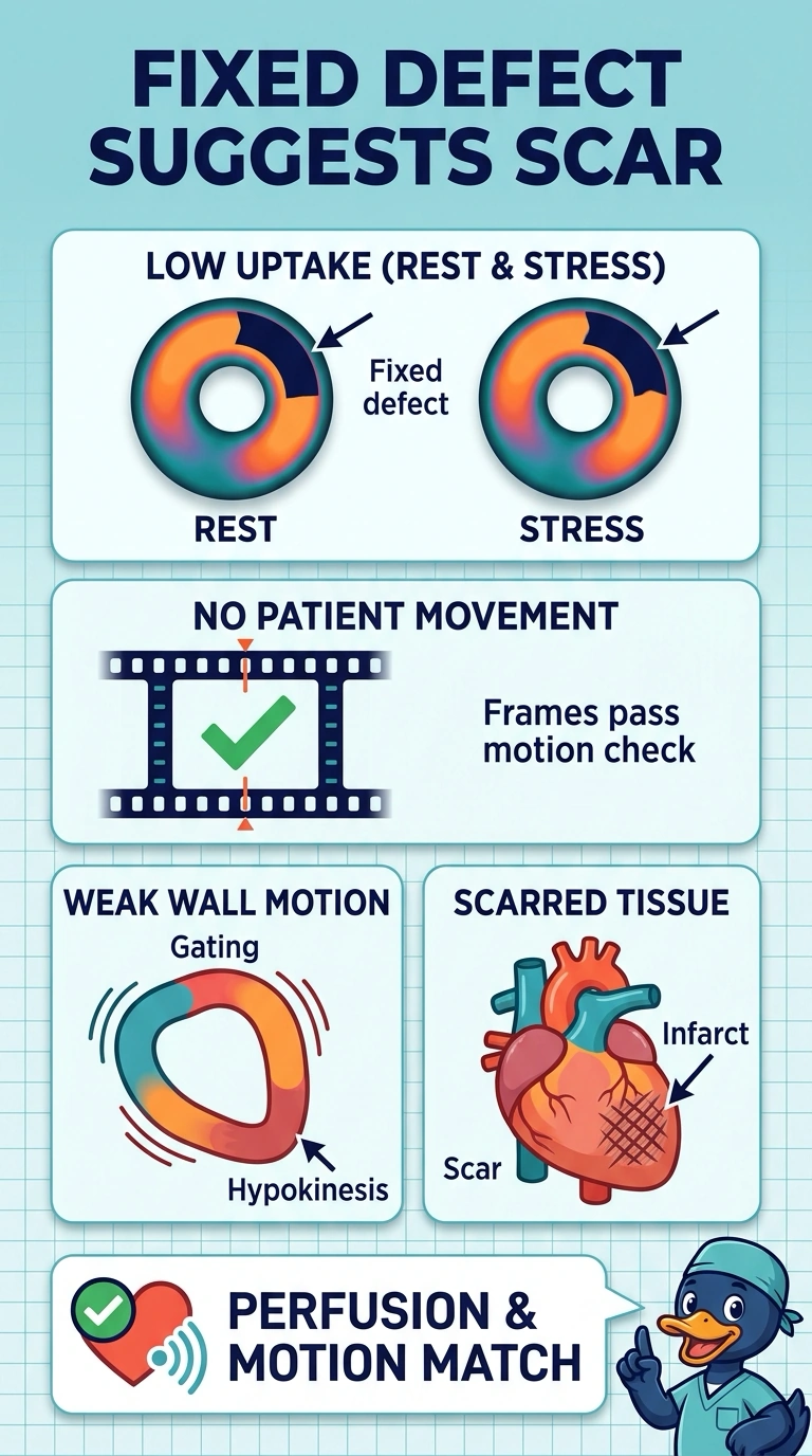
On a rest and stress myocardial perfusion study, the inferior wall shows reduced tracer uptake on both acquisitions, all frames passed the motion check, and the gated frames show that same segment contracting poorly. Which pathophysiologic explanation best accounts for this pattern?
Select an answer to reveal the explanation.
Short Explanation
When you see low uptake and weak contraction in the same segment on both rest and stress images, an infarct scar is the best fit—it’s like a patch that takes up less tracer and moves less. Since your motion checks passed and attenuation alone wouldn’t weaken contraction, this fixed defect with hypokinesis points to scar, though it isn’t definitive by itself.
Full Explanation
A fixed perfusion defect paired with a matching wall-motion abnormality is suggestive of infarct scar. Perfusion-tracer uptake reflects regional blood flow and viable myocardial tissue; infarcted tissue may show reduced uptake and contractility. Attenuation can reduce apparent inferior-wall uptake without causing a matching contraction abnormality. Reversible ischemia typically produces reduced stress uptake with improvement at rest, unlike the pattern described. Passing a motion check reduces concern for motion artifact but does not exclude every source of image artifact. Interpret gated function together with perfusion images and other clinical findings; the combination favors scar but is not by itself definitive.